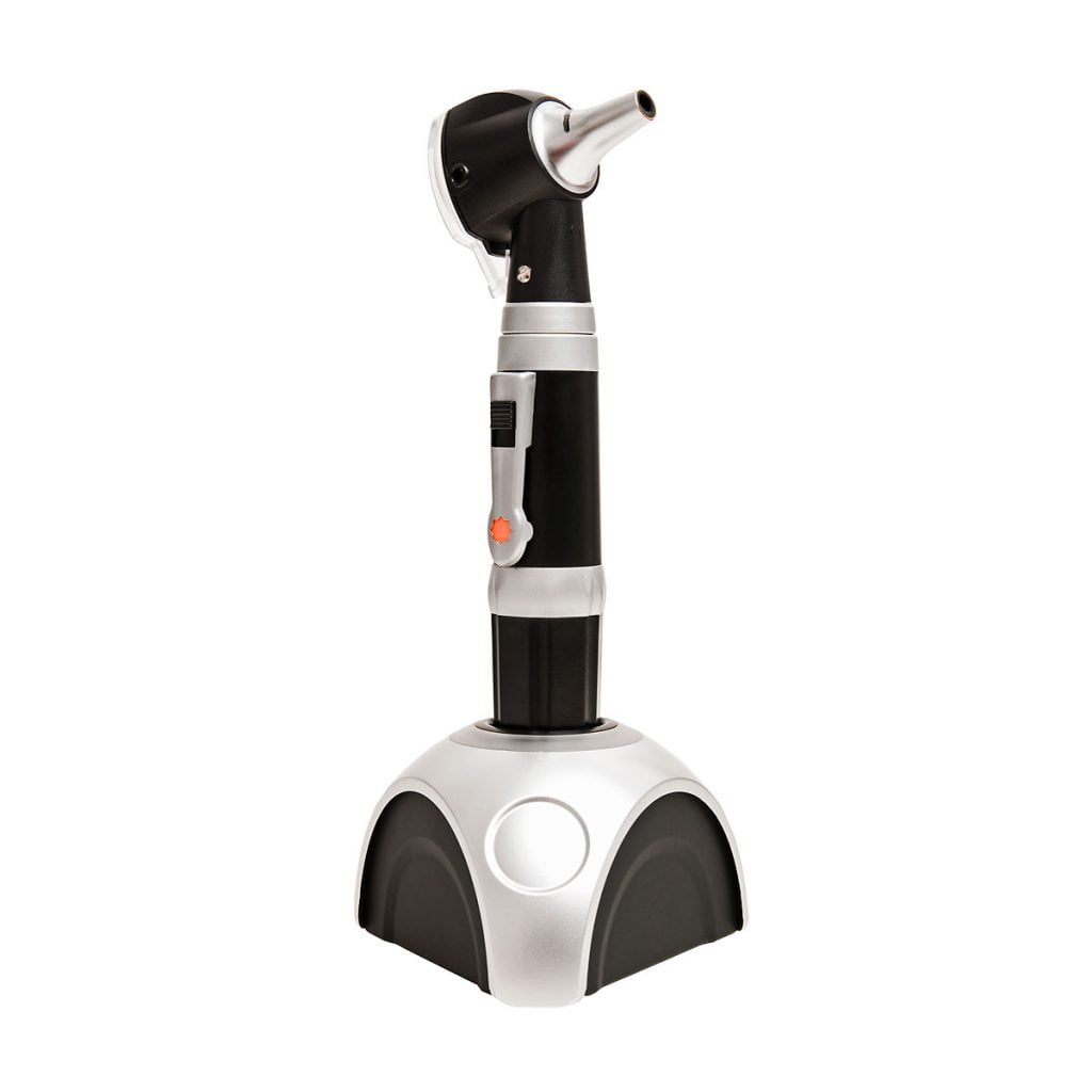
Fiber optic otoscopes are medical instruments used to examine the ear canal and eardrum. They use a fiber optic light source to provide bright, clear illumination of the ear canal, allowing healthcare professionals to see any abnormalities or conditions within the ear.
They typically consist of a handle with a battery-powered light source, a speculum (a cone-shaped attachment for the end of the otoscope), and a series of fibre optic cables that transmit the light from the handle to the speculum.
The use of fibre optics allows for a bright, focused beam of light to illuminate the ear canal without producing excessive heat, which can be uncomfortable for patients.
Fibre optic otoscopes are commonly used by healthcare professionals, including doctors, nurses, and audiologists, to diagnose ear infections, assess hearing loss, and monitor the healing process after surgery or treatment. They are an essential tool in the diagnosis and treatment of many ear-related conditions and are generally considered to be safe, effective, and reliable.
What Are They Used For?
- Ear infections: Fiber optic otoscopes are used to examine the ear canal and eardrum for signs of infection, including redness, swelling, and discharge. This allows healthcare professionals to diagnose and treat ear infections, which can be caused by bacteria, viruses, or fungi.
- Hearing loss: Fiber optic otoscopes can help diagnose hearing loss by examining the eardrum and the structures of the middle ear. This allows healthcare professionals to identify any abnormalities that may be causing hearing loss and recommend the appropriate treatment.
- Foreign objects: Fiber optic otoscopes can be used to locate and remove foreign objects that may have become lodged in the ear canal, such as small toys or insects.
- Wax buildup: Fiber optic otoscopes can help identify and remove excessive earwax that may be blocking the ear canal and affecting hearing.
- Surgery and treatment: Fiber optic otoscopes are often used to monitor the healing process after surgery or treatment for ear-related conditions, such as a perforated eardrum or a cholesteatoma.
Overall, fibre optic otoscopes are a crucial tool in the diagnosis and treatment of many ear-related conditions and play an important role in maintaining ear health.
How Does Fibre Optic Imaging Work?
Fiber optic imaging is a technology that uses fiber optic cables to transmit images from one location to another. The process involves the use of a light source, a fiber optic cable, and an imaging device.
Here is a general overview of how fiber optic imaging works:
Light source: A light source, such as a high-intensity LED or laser, produces light that is focused into a fiber optic cable.
Fiber optic cable: The fiber optic cable is a thin, flexible strand made of glass or plastic fibers. The light travels through the cable by reflecting off the walls of the fibers and is transmitted along the length of the cable.
Imaging device: At the other end of the fiber optic cable, an imaging device, such as a camera or microscope, captures the transmitted light and produces an image.
In medical applications, fiber optic imaging is commonly used to examine the inside of the body, such as the gastrointestinal tract or the urinary system. In this case, a fiber optic cable with a tiny camera on the end is inserted into the body through a small incision or natural opening. The camera captures images of the internal organs or tissues, which are transmitted back through the fiber optic cable to a monitor where they can be viewed by a healthcare professional.
Fibre optic imaging is also used in many other fields, such as telecommunications, engineering, and aerospace, where the ability to transmit images over long distances without loss of quality is important.


